
How is low testosterone diagnosed? What the guidelines and studies show
Guidelines diagnose low testosterone only when symptoms or signs are matched by low results on two morning blood tests. Testosterone changes with the time of day, after a meal and between laboratories, and further tests show whether the cause lies in the testes or in the pituitary.
The grade ModerateDiagnosing low testosterone with symptoms, two morning total testosterone tests and targeted follow-up tests
In brief. The two main US guidelines diagnose low testosterone only in men who have symptoms or signs of deficiency and low total testosterone on at least two separate morning blood tests.12 The Endocrine Society uses 264 ng/dL (9.2 nmol/L), the lower limit of a harmonized range in healthy nonobese young men, for laboratories whose assays are certified by the US Centers for Disease Control and Prevention (CDC). The American Urological Association (AUA) uses a total testosterone below 300 ng/dL.142 Testosterone is higher in the morning than the afternoon, varies from day to day and falls after a glucose drink, so a single result misclassifies many men.567 Unbound testosterone, the small fraction not bound to proteins in the blood, is measured when sex hormone-binding globulin (SHBG) is abnormal or total testosterone is borderline. Luteinizing hormone (LH) and follicle-stimulating hormone (FSH) then show whether the problem lies in the testes or in the pituitary and hypothalamus.1 The Endocrine Society recommends against screening the general population, and neither guideline uses saliva tests.12
A testosterone level is measured with a blood test and compared with a cutoff. The comparison is harder than it looks. The level changes through the day, laboratory methods disagree, most testosterone in blood is bound to proteins whose concentrations differ between men, and the symptoms attributed to low testosterone are common in men whose levels are normal. This article describes how the Endocrine Society’s 2018 guideline and the AUA’s 2018 guideline approach the diagnosis, and the studies behind their advice. The first article in this series covers what treatment does, and the second covers heart, prostate and blood safety and the monitoring guidelines describe.
What do the guidelines say about diagnosing low testosterone?
The Endocrine Society recommends diagnosing hypogonadism only in men with symptoms and signs of testosterone deficiency and testosterone concentrations that are “unequivocally and consistently low,” a strong recommendation based on moderate-quality evidence. It recommends a fasting morning total testosterone as the first test, measured with an accurate and reliable assay, and repeating the measurement to confirm the diagnosis.1 Its task force rated the quality of the evidence for each recommendation, and six of its ten members, including the chair, declared research support from or consulting for drug companies, among them AbbVie, Clarus Therapeutics, Lipocine, Bayer and Besins, which make or develop testosterone products.1
The AUA guideline makes three linked statements. Clinicians should use a total testosterone below 300 ng/dL as “a reasonable cut-off” (moderate recommendation, evidence grade B). The diagnosis should be made only after two total testosterone measurements taken on separate occasions, both in the early morning (strong recommendation, grade A). And the clinical diagnosis is made only when low levels are combined with symptoms or signs (moderate recommendation, grade B).2 The panel chose 300 ng/dL because randomized trials of treatment had enrolled men with levels below 350 ng/dL and a median baseline of 249 ng/dL, and it noted that other societies had used thresholds from 230 to 350 ng/dL.2 The guideline’s chair disclosed consulting for Pfizer and Lilly, and the AUA confirmed the guideline’s validity on review in 2024.23
Why does the time of day matter?
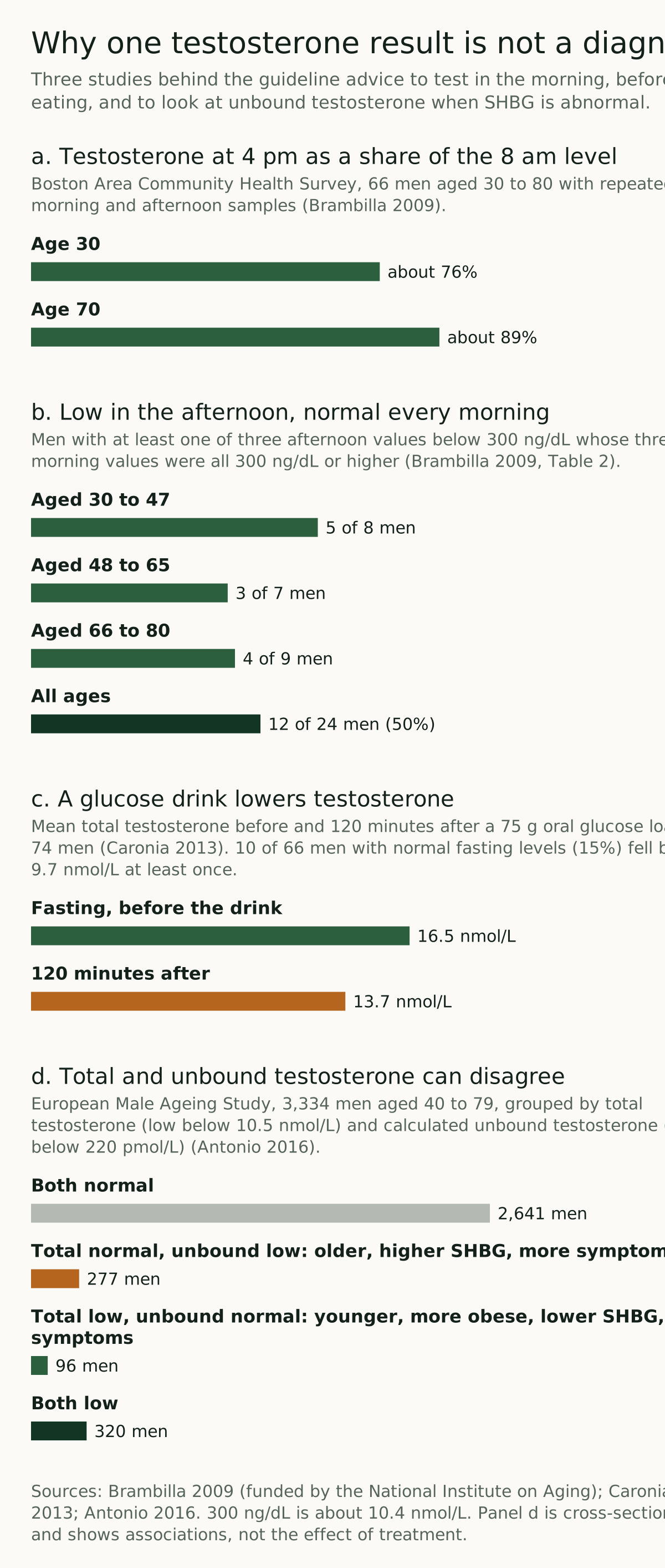
Testosterone is highest in the morning. In a study of 66 randomly selected men aged 30 to 80 from the Boston Area Community Health Survey, each gave blood at three morning and three afternoon visits. At age 30, testosterone at 4 pm averaged about 76% of the level at 8 am. At age 70 it averaged about 89%, because the daily rhythm flattens with age.5 Among the 62 men with complete sets of samples, 24 had at least one afternoon value below 300 ng/dL, and 12 of them had normal values at all three morning visits. The pattern held in each age group, including 4 of 9 men aged 66 to 80.5 The study was funded by the National Institute on Aging.5
Neither guideline names a clock time. The Endocrine Society says morning and the AUA says early morning.12 One group notes that reference values are generally based on samples drawn between 7 and 10 am.8 Both guidelines also advise against testing during or soon after an acute illness. The Endocrine Society adds short-term use of medicines that suppress testosterone, such as opioids, and the AUA cites a small study in which acute respiratory infections lowered mean testosterone by 10%.12
Why is a second test needed?
Testosterone also varies from one day to the next. In 132 men aged 30 to 79 from the same Boston survey, paired blood samples were drawn 1 to 3 days apart at entry and again at 3 and 6 months. Biological variation accounted for more of the differences than laboratory error. Across the hormones studied, two measurements in the same man differed by more than 18% to 28% about half the time and by more than 27% to 54% about a quarter of the time. The authors concluded that one sample is generally not enough and that more than three is probably not needed.6 The Endocrine Society states that 30% of men with an initial testosterone in the hypogonadal range have a normal level when it is measured again, and that at least two measurements are needed to diagnose deficiency with confidence.1
The AUA advises using the same laboratory and the same method for repeat tests. If the first result is below 300 ng/dL and the second is normal, it leaves a third test to the clinician’s judgment, and it notes that no evidence defines the best interval between tests.2
Does eating before the test matter?
The two guidelines differ here. The Endocrine Society recommends measuring testosterone in the morning after an overnight fast, because glucose and food intake suppress it.1 In a study of 74 men aged 19 to 74 who drank 75 g of glucose, mean testosterone fell by 25% and was still lower at 120 minutes than at baseline (13.7 versus 16.5 nmol/L). Of 66 men with normal fasting levels, 10 (15%) fell into the hypogonadal range at least once. LH and SHBG did not change.7 A Swedish study found testosterone 30% lower 60 to 120 minutes after a 550-kcal meal than after an overnight fast.8 The AUA describes this evidence as particularly weak and does not recommend that clinicians insist on fasting.2
Does the laboratory matter?
It can matter a great deal. Total testosterone is measured by immunoassay or by liquid chromatography with tandem mass spectrometry. When 1,133 laboratories using 14 assays measured the same quality-control sample from one hypogonadal man, the results ranged from 45 to 365 ng/dL.1 Some immunoassays are also affected by biotin, and the Endocrine Society advises stopping biotin supplements for at least 72 hours before LH and FSH tests, which are susceptible to the same interference.1 An earlier Endocrine Society position statement called for laboratory proficiency testing based on accuracy against samples of known concentration, not only agreement with other laboratories using the same method.9
Since 2010 the CDC has run a testosterone standardization program. Laboratories and assay makers are certified when their results stay within a mean bias of ±6.4% of the CDC reference method over 2.50 to 1,000 ng/dL.110 A harmonization study calibrated the assays of four cohorts in the United States and Europe, with 9,054 men in total, against the CDC method. In healthy nonobese men aged 19 to 39, the harmonized 2.5th and 97.5th percentiles were 264 and 916 ng/dL, and the 5th and 95th percentiles 303 and 852 ng/dL.4 The study was publicly funded, and its senior author, who also chaired the Endocrine Society task force, disclosed grants from and consulting for AbbVie.4 The Endocrine Society says this range can be used for CDC-certified assays, and that ranges from laboratories outside an accuracy-based program may not identify hypogonadism accurately.1 The AUA gives an example: a result of 250 ng/dL may be marked normal by a laboratory and yet fall below the guideline cutoff, and in that case it advises using the absolute value.2
What is SHBG, and when does unbound testosterone matter?
Most testosterone in blood is bound to proteins. The AUA guideline gives the shares as about 50% bound loosely to albumin, 44% bound tightly to SHBG and 4% to cortisol-binding globulin, leaving about 2% unbound.2 The Endocrine Society gives the unbound share as 2.0% to 4.0%.1 Unbound testosterone is the fraction not attached to any protein, and “bioavailable” testosterone is the unbound fraction plus the portion loosely bound to albumin.1
Because total testosterone includes the SHBG-bound portion, conditions that change SHBG move the total without necessarily changing the unbound level. The Endocrine Society lists obesity, diabetes, glucocorticoids, some progestins, androgenic steroids, nephrotic syndrome, hypothyroidism and acromegaly as causes of low SHBG. It lists aging, HIV infection, cirrhosis and hepatitis, hyperthyroidism, some anticonvulsants and estrogens as causes of high SHBG, and variants in the SHBG gene can push it either way.1 Low SHBG can bring total testosterone below the normal range while the unbound level stays normal. High SHBG can raise total testosterone well above 400 ng/dL even when the unbound level is low.1
The Endocrine Society recommends measuring unbound testosterone in men with these conditions and in men whose total testosterone is near the lower limit, giving 200 to 400 ng/dL as an example. It says the measurement is generally unnecessary when total testosterone is far below the limit, such as under 150 ng/dL.1 The reference method is equilibrium dialysis. The alternative is to calculate the unbound level from total testosterone, SHBG and albumin, and in a 1999 comparison calculated values were almost identical to dialysis values except in pregnancy.111 Direct “analog” immunoassays sold for unbound testosterone gave values that were only a fraction of the dialysis result, and the Endocrine Society says they should not be used.111 No harmonized reference range exists for unbound testosterone, so laboratories set their own.1
The AUA takes a more limited view. It does not recommend unbound testosterone as the primary diagnostic test, because calculated values depend on the accuracy of the total testosterone, SHBG and albumin results. It cites an analysis of 3,219 men in which the unbound level added nothing when total testosterone was below 231 ng/dL and might help when it was between 230 and 317 ng/dL.2
The strongest population data come from the European Male Ageing Study (EMAS), which enrolled community-dwelling men aged 40 to 79 in eight European centers and was funded by the European Commission.415 Of 3,334 men, 277 had normal total but low calculated unbound testosterone. They were older and in poorer health, had higher SHBG and LH, and reported more sexual and physical symptoms. Another 96 men had low total but normal unbound testosterone. They were younger and more obese, had lower SHBG and normal LH, and lacked features of deficiency.12 These are associations measured at one point in time, not evidence that testing unbound testosterone improves the results of treatment.
How do LH and FSH separate primary from secondary hypogonadism?
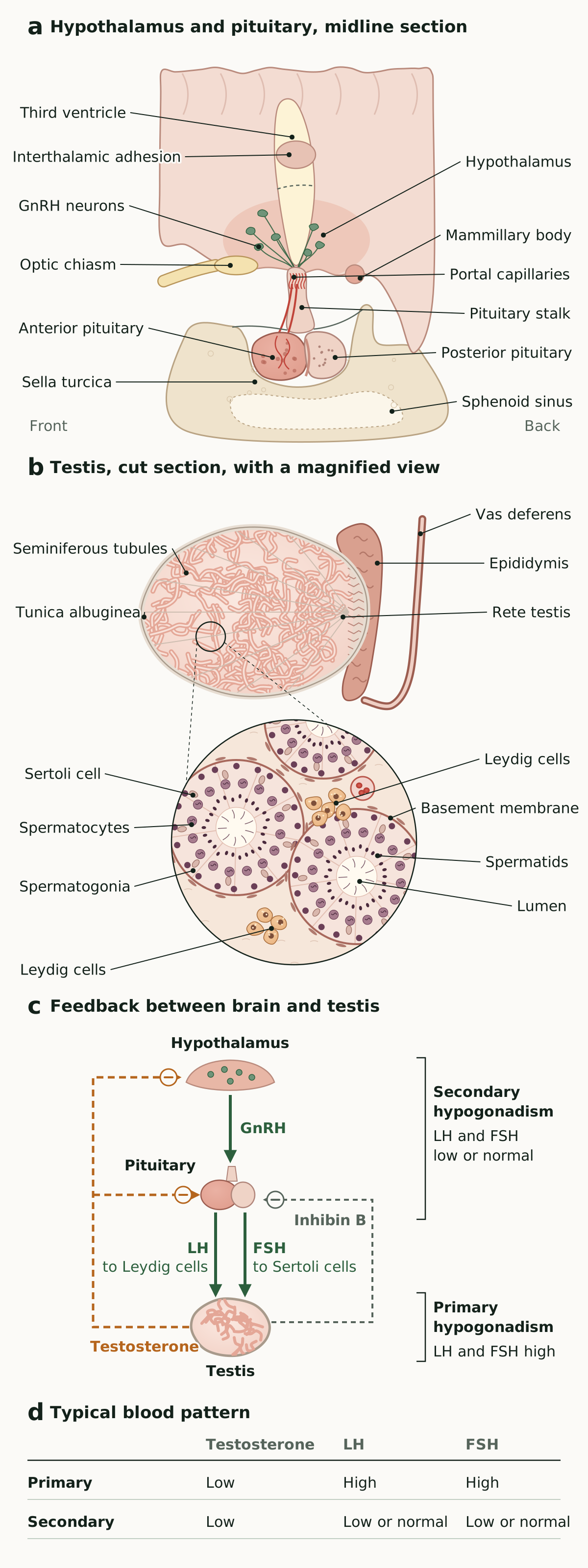
The testes are controlled from the brain. Gonadotropin-releasing hormone (GnRH) from the hypothalamus stimulates the anterior pituitary to release LH and FSH. LH drives the Leydig cells between the seminiferous tubules to make testosterone, and FSH acts on the Sertoli cells inside the tubules that support sperm production. Testosterone, and inhibin B from the Sertoli cells, feed back to restrain GnRH and gonadotropin secretion.132
Primary hypogonadism starts in the testes. Testosterone is low and LH and FSH are high, because the pituitary is responding to the lack of feedback. Causes listed by the Endocrine Society include Klinefelter syndrome, undescended testes, some cancer chemotherapy, radiation to the testes, trauma, torsion, orchitis, HIV infection and myotonic dystrophy. Secondary hypogonadism starts in the hypothalamus or pituitary, with low testosterone and low or “inappropriately normal” LH and FSH. Its causes include high prolactin, severe obesity, iron overload, opioids, glucocorticoids, withdrawal from anabolic steroids, pituitary or hypothalamic tumors, head trauma and pituitary surgery or radiation.1 Both guidelines recommend measuring LH once testosterone is confirmed low. The Endocrine Society adds FSH, a strong recommendation based on moderate-quality evidence.12 The distinction matters for fertility, because sperm production can be restored with gonadotropin treatment in secondary hypogonadism but not in primary hypogonadism.1
What other tests do guidelines describe?
For secondary hypogonadism, the Endocrine Society says clinicians should measure prolactin and iron saturation and, in certain cases, test other pituitary hormones and image the pituitary with MRI. It suggests MRI when testosterone is below 150 ng/dL, when several pituitary hormones are deficient, when prolactin stays high, or when there are signs of a pituitary mass such as headache or loss of vision. For primary hypogonadism of unknown cause, especially with testes smaller than 6 mL, it recommends a karyotype to look for Klinefelter syndrome.1 The AUA recommends measuring prolactin when LH is low or low-normal and referring men with persistently high prolactin of unknown cause for endocrine evaluation. It also recommends estradiol for men with breast symptoms or breast enlargement, a reproductive evaluation before treatment for men interested in fertility, and hemoglobin, hematocrit and, in men over 40, PSA before treatment.2
The yield of pituitary imaging is modest and its rules are imperfect. In an Edinburgh series of 281 men with low testosterone, normal prolactin and low or normal gonadotropins who had pituitary MRI, 83.6% of scans were normal, 8.5% showed microadenomas, 1.8% macroadenomas and 0.4% a craniopharyngioma. The Endocrine Society’s criteria would have prompted imaging in only 3 of the 6 men with significant pituitary disease.14
Many causes of secondary hypogonadism are functional. The Endocrine Society notes that obesity, opioid use and systemic illness can suppress testosterone and that some of these may be reversible by managing the underlying condition.1 Any change to a prescribed medicine, including opioids or glucocorticoids, is a decision for the prescribing clinician.
Which symptoms point to low testosterone?
Few symptoms are specific. The Endocrine Society lists incomplete or delayed sexual development, loss of body hair and very small testes (under 6 mL) as specific. It lists reduced libido, fewer spontaneous erections or erectile dysfunction, breast discomfort or enlargement, infertility, low bone density or low-trauma fracture and hot flushes as suggestive. Low energy, low mood, poor concentration, sleep disturbance, mild unexplained anemia, reduced muscle bulk and increased body fat are listed as nonspecific.1
Population studies show how loose the link is. EMAS surveyed 3,369 men aged 40 to 79 and measured total testosterone by mass spectrometry in morning samples. Poor morning erections, low sexual desire, erectile dysfunction, inability to do vigorous activity, depression and fatigue were all related to testosterone, but only the three sexual symptoms had a syndromic association with low levels. The investigators proposed defining late-onset hypogonadism as at least three sexual symptoms with total testosterone below 11 nmol/L (about 317 ng/dL) and calculated unbound testosterone below 220 pmol/L (64 pg/mL).15 In the Boston Area Community Health Survey of 1,475 men aged 30 to 79, about 24% had total testosterone below 300 ng/dL, but many men with low levels had no symptoms, including 47.6% of those aged 50 or older. Low testosterone with symptoms was found in 5.6% overall.16 Both guidelines advise against relying on symptom questionnaires to decide who has deficiency.12
How much does testosterone fall with age?
Testosterone declines gradually with age, and other conditions account for part of the decline. In the Baltimore Longitudinal Study of Aging, stored samples from 890 men showed total testosterone falling by an average of 0.124 nmol/L per year. Using total testosterone criteria, about 20% of men over 60, 30% over 70 and 50% over 80 had values in the hypogonadal range.17 In a cross-sectional analysis of 3,200 EMAS men, age was linked to lower unbound testosterone with higher LH, a testicular pattern. Obesity was linked to lower total testosterone, by 5.09 nmol/L at a body mass index of 30 or more, with LH unchanged or lower, a central pattern.18 EMAS then classified men by testosterone and LH. Secondary hypogonadism was found in 11.8%, primary in 2.0% and “compensated” hypogonadism, with normal testosterone and high LH, in 9.5%. Obesity was strongly associated with the secondary pattern and older age with the primary and compensated patterns.19 The harmonization study also published age-specific ranges for nonobese men by decade of age.4 The Endocrine Society bases its lower limit on healthy nonobese young men.1
Should all men be tested for low testosterone?
The Endocrine Society recommends against routine screening of men in the general population, a strong recommendation based on low-quality evidence. It notes that no clinical trials have tested a screening strategy, that the benefits and risks of long-term treatment in men without symptoms are unclear, and that low levels often occur without symptoms. It places a high value on avoiding labeling, testing, treating and monitoring healthy men.1 It suggests testing men with conditions in which low testosterone is common, including pituitary disease or radiation, treatment with opioids or glucocorticoids, withdrawal from anabolic steroids, HIV-associated weight loss, infertility, osteoporosis or low-trauma fracture, and low libido or erectile dysfunction.1 The AUA says clinicians should consider measuring total testosterone even without symptoms or signs in men with unexplained anemia, bone density loss, diabetes, exposure to chemotherapy or testicular radiation, HIV/AIDS, chronic narcotic use, male infertility, pituitary dysfunction or chronic corticosteroid use.2
A 2024 review in the Journal of the Endocrine Society described “low T” as frequently diagnosed in men’s health clinics and other venues without actual hormone testing or further evaluation, and often treated with doses above the physiological range that suppress the body’s own testosterone and sperm production.20
What about saliva and at-home tests?
Neither guideline uses saliva tests. The Endocrine Society’s recommendations, and its 264 ng/dL lower limit, apply to serum total testosterone measured with an accurate assay, preferably one certified by the CDC.14 The AUA’s table of assays lists salivary testosterone as simple and accessible and as correlating with calculated unbound testosterone in serum. It also lists it as not FDA approved, as needing extensive, highly skilled sample preparation, and as raising concerns about specimen stability.2 Salivary testosterone has been measured by mass spectrometry in 1,675 men in a British population survey, where the mean fell from 322.6 pmol/L at age 18 to 153.9 pmol/L at age 69. The authors presented these distributions as a resource for population research.21 A 2025 comparison in 218 young adults, 47 of them men, found that a commercial salivary immunoassay performed poorly against mass spectrometry, though testosterone fared better than estradiol and progesterone.22 Neither guideline addresses home-collected blood samples, and their cutoffs were derived from laboratory serum measurements.12
Who should not start testosterone, and how is it monitored?
Testosterone is a prescription medicine, and whether to start, adjust or stop it is a decision for a man and his clinician. A diagnosis does not settle whether treatment is appropriate. The Endocrine Society recommends against testosterone in men planning fertility in the near term and in men with breast or prostate cancer, a palpable prostate nodule or induration, a PSA above 4 ng/mL (or above 3 ng/mL with high prostate cancer risk) without further urological evaluation, an elevated hematocrit, untreated severe obstructive sleep apnea, severe lower urinary tract symptoms, uncontrolled heart failure, a heart attack or stroke within the last 6 months, or thrombophilia.1 The AUA advises against testosterone in men trying to conceive and against starting it for three to six months after a cardiovascular event.2
During treatment, both guidelines call for repeat testosterone levels, hematocrit and, in the relevant age groups, PSA, with defined points at which to act. These plans are described in the second article in this series.12
How good is the evidence, and who paid for it?
Most of the diagnostic advice rests on measurement studies and population cohorts, not on trials. The studies of daily and day-to-day variation were small (66 and 132 men) and drawn from one Boston survey.56 The meal studies were small and short.78 EMAS and the Boston survey are large population samples, but their analyses of symptoms and unbound testosterone are cross-sectional.151216 The harmonized range rests on four cohorts calibrated to a reference method.4 No trial has compared diagnostic strategies, and none has tested screening.1
The cohort and measurement studies whose funding we could check were publicly funded: the daily-variation study by the National Institute on Aging, EMAS by the European Commission, and the harmonization study by US and European public grants.54 Industry ties appear mainly at the guideline level. Six of the ten Endocrine Society task force members declared relationships with companies, including makers of testosterone products, and the AUA panel chair disclosed consulting for Pfizer and Lilly.12
What remains uncertain?
The right cutoff is still debated. The guidelines use 264 and 300 ng/dL, the EMAS definition corresponds to 11 nmol/L, and the AUA notes that societies have used values from 230 to 350 ng/dL.1215 There is no harmonized reference range for unbound testosterone, and the guidelines disagree on how central it should be.12 The best interval between repeat tests is unknown, and the guidelines differ on fasting.21 Neither guideline uses age-specific ranges, although the harmonization study published them.4 The criteria for pituitary imaging miss some men with pituitary disease.14
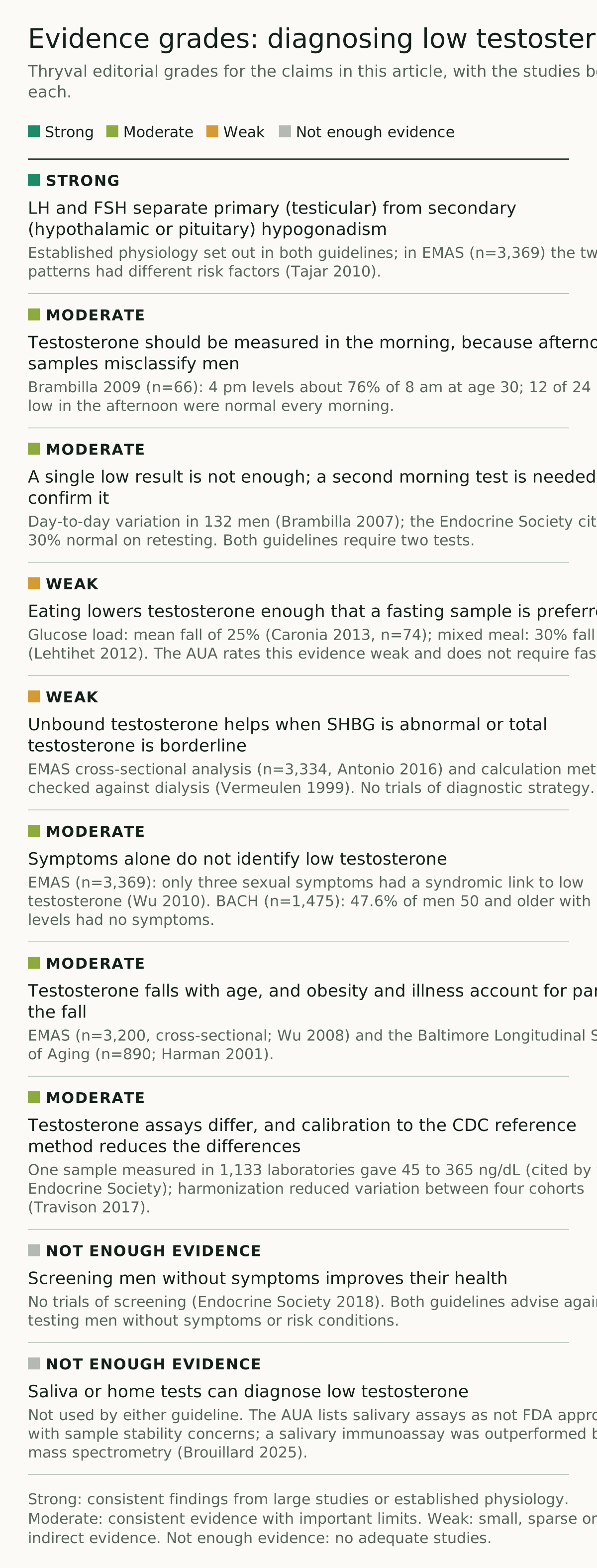
Evidence grades
The grades below are Thryval editorial judgments based on the studies cited in this article. The grading rules are set out on our Research page.
Frequently asked questions
What is a normal testosterone level for men?
The Endocrine Society gives 264 to 916 ng/dL as the harmonized range in healthy nonobese men aged 19 to 39, for CDC-certified assays.14 The AUA treats a total testosterone below 300 ng/dL as low.2 Laboratory ranges can differ from both.
What time of day should testosterone be tested?
In the morning. In one study, testosterone at 4 pm averaged about 76% of the 8 am level in 30-year-old men, and half the men with a low afternoon value had normal values on every morning visit.5 The AUA recommends two early-morning tests on separate days.2
Do I need to fast before a testosterone blood test?
The Endocrine Society recommends a fasting morning sample, because a glucose drink lowered mean testosterone by 25% in one study.17 The AUA considers the evidence weak and does not insist on fasting.2 The ordering clinician or laboratory will give instructions.
What is SHBG, and why does it affect testosterone results?
SHBG is a blood protein that binds testosterone tightly. Obesity and diabetes lower it, and aging, liver disease and some anticonvulsants raise it, which moves total testosterone up or down without the same change in unbound testosterone. In those cases the Endocrine Society recommends measuring or calculating unbound testosterone.1
What is the difference between primary and secondary hypogonadism?
Primary hypogonadism starts in the testes, so LH and FSH are high. Secondary hypogonadism starts in the pituitary or hypothalamus, so LH and FSH are low or normal. Secondary hypogonadism prompts tests such as prolactin and iron saturation, and sometimes a pituitary MRI.12
Are saliva or at-home testosterone tests accurate?
Neither major US guideline uses saliva tests, and the AUA lists salivary assays as not FDA approved, with concerns about sample stability.2 The guideline cutoffs apply to blood samples measured in a laboratory with an accurate assay.1
Implications for patients and clinicians
A diagnosis of low testosterone, as the guidelines define it, needs symptoms or signs and low results on two morning blood tests, preferably from a laboratory with a CDC-certified assay.12 When SHBG is likely to be abnormal or the total is borderline, unbound testosterone can clarify the result. LH and FSH, and in secondary hypogonadism prolactin and iron studies, point toward the cause.1 A single afternoon or nonfasting result, a symptom questionnaire or a saliva test is not a basis for diagnosis under either guideline.512 Treatment decisions, including the contraindications and monitoring described above, belong with the treating clinician.
This article is for education and is not medical advice.
References
-
Bhasin S, Brito JP, Cunningham GR, et al. Testosterone therapy in men with hypogonadism: an Endocrine Society clinical practice guideline. J Clin Endocrinol Metab. 2018;103(5):1715–1744. Clinical practice guideline using GRADE methods; diagnostic recommendations 1.1 to 1.4, Tables 1 to 4 and Figure 1; task force conflicts listed in the appendix. PubMed 29562364 ↩↩↩↩↩↩↩↩↩↩↩↩↩↩↩↩↩↩↩↩↩↩↩↩↩↩↩↩↩↩↩↩↩↩↩↩↩↩↩↩↩↩↩↩↩↩↩↩↩↩↩↩↩
-
Mulhall JP, Trost LW, Brannigan RE, et al. Evaluation and management of testosterone deficiency: AUA guideline. J Urol. 2018;200(2):423–432. Clinical guideline based on a systematic review; guideline statements 1 to 12 and Table 4 (assays); panel disclosures in the full text. PubMed 29601923 · AUA guideline PDF ↩↩↩↩↩↩↩↩↩↩↩↩↩↩↩↩↩↩↩↩↩↩↩↩↩↩↩↩↩↩↩↩↩↩↩↩↩↩↩
-
American Urological Association. Testosterone deficiency guideline (published 2018, reviewed and validity confirmed 2024). AUA guideline page ↩
-
Travison TG, Vesper HW, Orwoll E, et al. Harmonized reference ranges for circulating testosterone levels in men of four cohort studies in the United States and Europe. J Clin Endocrinol Metab. 2017;102(4):1161–1173. Cross-calibration of four cohorts, n=9054; funded by US and European public grants; the senior author disclosed AbbVie grants and consulting. PMC5460736 · PubMed 28324103 ↩↩↩↩↩↩↩↩↩↩↩
-
Brambilla DJ, Matsumoto AM, Araujo AB, McKinlay JB. The effect of diurnal variation on clinical measurement of serum testosterone and other sex hormone levels in men. J Clin Endocrinol Metab. 2009;94(3):907–913. Repeated-sampling study, n=66; funded by the National Institute on Aging; one author reported consulting for drug companies. PMC2681273 · PubMed 19088162 ↩↩↩↩↩↩↩↩↩
-
Brambilla DJ, O’Donnell AB, Matsumoto AM, McKinlay JB. Intraindividual variation in levels of serum testosterone and other reproductive and adrenal hormones in men. Clin Endocrinol (Oxf). 2007;67(6):853–862. Repeated-sampling study, n=132. PubMed 18052942 ↩↩↩↩
-
Caronia LM, Dwyer AA, Hayden D, et al. Abrupt decrease in serum testosterone levels after an oral glucose load in men: implications for screening for hypogonadism. Clin Endocrinol (Oxf). 2013;78(2):291–296. Cross-sectional study with oral glucose tolerance testing, n=74. PubMed 22804876 ↩↩↩↩↩
-
Lehtihet M, Arver S, Bartuseviciene I, Pousette A. S-testosterone decrease after a mixed meal in healthy men independent of SHBG and gonadotrophin levels. Andrologia. 2012;44(6):405–410. Fasting versus standard-meal sampling study in healthy men. PubMed 22524522 ↩↩↩↩
-
Rosner W, Auchus RJ, Azziz R, Sluss PM, Raff H. Position statement: utility, limitations, and pitfalls in measuring testosterone: an Endocrine Society position statement. J Clin Endocrinol Metab. 2007;92(2):405–413. Expert position statement. PubMed 17090633 ↩
-
US Centers for Disease Control and Prevention. Hormones certified assays and participants (CDC Hormone Standardization Program), updated Oct 6, 2026. Accessed Nov 11, 2026. CDC page ↩
-
Vermeulen A, Verdonck L, Kaufman JM. J Clin Endocrinol Metab. 1999;84(10):3666–3672. Laboratory method comparison of equilibrium dialysis, calculated values and direct analog immunoassay across a wide range of SHBG concentrations. PubMed 10523012 ↩↩↩
-
Antonio L, Wu FCW, O’Neill TW, et al; European Male Ageing Study Group. J Clin Endocrinol Metab. 2016;101(7):2647–2657. Cross-sectional analysis of the European Male Ageing Study comparing low total and low calculated unbound testosterone, n=3334. PubMed 26909800 ↩↩↩
-
Oduwole OO, Huhtaniemi IT, Misrahi M. The roles of luteinizing hormone, follicle-stimulating hormone and testosterone in spermatogenesis and folliculogenesis revisited. Int J Mol Sci. 2021;22(23):12735. Narrative review; the authors declared no conflicts of interest. PMC8658012 · PubMed 34884539 ↩↩
-
Dalvi M, Walker BR, Strachan MW, Zammitt NN, Gibb FW. The prevalence of structural pituitary abnormalities by MRI scanning in men presenting with isolated hypogonadotrophic hypogonadism. Clin Endocrinol (Oxf). 2016;84(6):858–861. Retrospective case series, n=281. PubMed 26733239 ↩↩
-
Wu FC, Tajar A, Beynon JM, et al; EMAS Group. Identification of late-onset hypogonadism in middle-aged and elderly men. N Engl J Med. 2010;363(2):123–135. Cross-sectional population survey, n=3369. PubMed 20554979 ↩↩↩↩↩
-
Araujo AB, Esche GR, Kupelian V, et al. Prevalence of symptomatic androgen deficiency in men. J Clin Endocrinol Metab. 2007;92(11):4241–4247. Population-based survey (Boston Area Community Health Survey), n=1475. PubMed 17698901 ↩↩↩
-
Harman SM, Metter EJ, Tobin JD, Pearson J, Blackman MR; Baltimore Longitudinal Study of Aging. J Clin Endocrinol Metab. 2001;86(2):724–731. Longitudinal cohort analysis of total testosterone and SHBG with age, n=890. PubMed 11158037 ↩↩
-
Wu FC, Tajar A, Pye SR, et al; European Male Aging Study Group. Hypothalamic-pituitary-testicular axis disruptions in older men are differentially linked to age and modifiable risk factors: the European Male Aging Study. J Clin Endocrinol Metab. 2008;93(7):2737–2745. Cross-sectional survey, n=3200. PubMed 18270261 ↩↩
-
Tajar A, Forti G, O’Neill TW, et al; EMAS Group. Characteristics of secondary, primary, and compensated hypogonadism in aging men: evidence from the European Male Ageing Study. J Clin Endocrinol Metab. 2010;95(4):1810–1818. Cross-sectional survey, n=3369. PubMed 20173018 ↩↩↩
-
McDermott MT. Pseudo-endocrine disorders: recognition, management, and action. J Endocr Soc. 2024;9(1):bvae226. Narrative review. PMC11694709 · PubMed 39749108 ↩
-
Keevil BG, Clifton S, Tanton C, et al. Distribution of salivary testosterone in men and women in a British general population-based sample: the Third National Survey of Sexual Attitudes and Lifestyles (Natsal-3). J Endocr Soc. 2017;1(1):14–25. Population survey with mass spectrometry, 1675 men. PMC5677216 · PubMed 29264442 ↩
-
Brouillard A, Davignon LM, Cernik R, et al. Comparing immunoassay and mass spectrometry techniques for salivary sex hormone analysis. Psychoneuroendocrinology. 2025;174:107379. Assay comparison in 218 young adults, 47 of them men; the authors declared no competing interests. PubMed 39933211 ↩↩